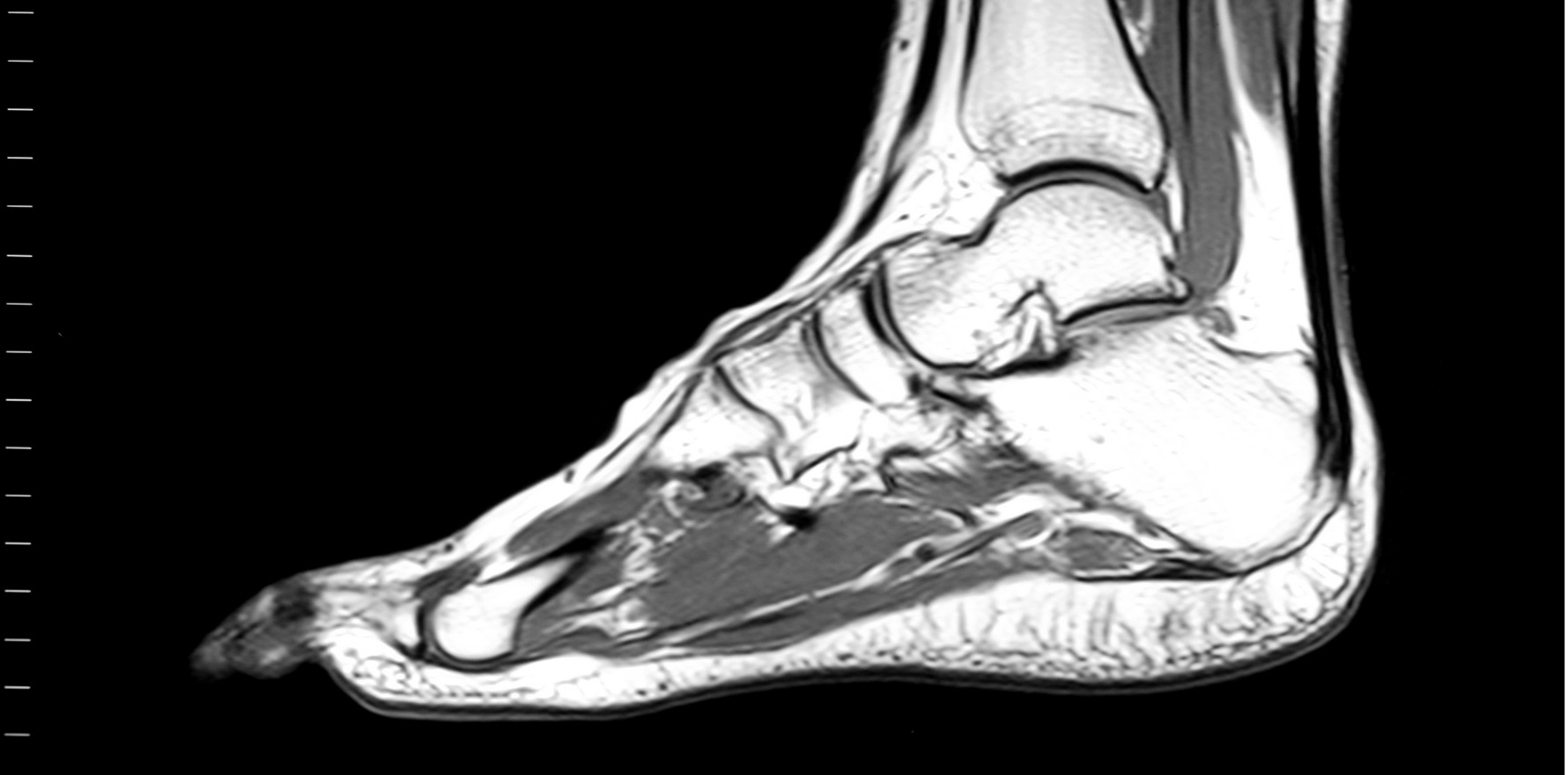
MRI-detected tenosynovitis predicts RA developing within a year, potentially preventing overtreatment.
MRI scans can help predict RA progression and could prevent overtreatment in select groups of patients with early arthritis, a large Dutch imaging study has found.
The prospective study of nearly 970 patients with undifferentiated arthritis (UA) tested the predictive value of MRI scans compared with the swollen joint counts, inflammatory markers and autoantibodies that are usually assessed.
The study, which was published in Rheumatology, found that detecting tenosynovitis on MRI predicted the patients more likely to develop RA and who would benefit from early treatment, before joint destruction occurs.
“The results of this study could be helpful in achieving precision medicine in patients with UA and in preventing overtreatment,” Dr Nikolet den Hollander and colleagues at the Leiden University Medical Center in the Netherlands wrote.
Two groups of early arthritis patients, who had at least one affected joint and symptoms for less than two years, were consecutively included over 10 years.
Patients either didn’t fulfill criteria for RA and had no clear alternative diagnosis, or their treating rheumatologist had indicated undifferentiated arthritis.
The predictive value of MRI was similar in both groups so MRI could be helpful in clinical practice and future imaging studies, the researchers wrote.
Contrast-enhanced MRI of the hands, wrist and feet taken at baseline were scored for inflammatory features, including bone inflammation, synovitis and tenosynovitis.
Of these inflammatory features, MRI-detected tenosynovitis was the strongest predictor of developing RA 12 months later. Patients with inflamed tendon sheaths on MRI were two-to-three times more likely to develop RA within a year, the study found.
MRI was most valuable among autoantibody-negative patients with oligoarthritis, where a negative MRI largely excluded the development of RA.
“The absence of MRI signs of tenosynovitis in this subset [of patients] predicts that RA is unlikely,” said Sydney-based radiologist Dr Sebastian Fung, who was not involved in the study.
This result may prevent overtreatment, the researchers said.
Moreover, MRI-detected tenosynovitis, but not synovitis or osteitis, was as predictive as the total inflammation score, suggesting that “in practice, only MRI-detected tenosynovitis can be assessed rather than evaluating all features”, Dr Hollander and colleagues wrote.
Professor Paul Bird, a rheumatologist at UNSW Sydney examining the use of MRI scans in inflammatory arthritis, said that based on the study results, MRI of the hands, wrists and feet could help classify patients at risk of developing RA and stratify treatment accordingly.
“One of the challenges for clinicians is classifying patients with ACPA-negative oligoarthritis and this can have implications for access to therapy,” Professor Bird said.
“The study provides evidence that MRI can be predictive of progression to RA in patients with undifferentiated arthritis,” he continued.
“Longer follow-up would be useful, but the results show utility of the method after 12 months.”


