
If we keep using our current MRI cut-offs, we could be wrongly diagnosing axial spondyloarthritis in healthy older people, a study finds
We’ve known for a little while that our MRI thresholds for axial spondyloarthritis diagnosis have a high false positive rate, but a German study has shown just how off the mark we are.
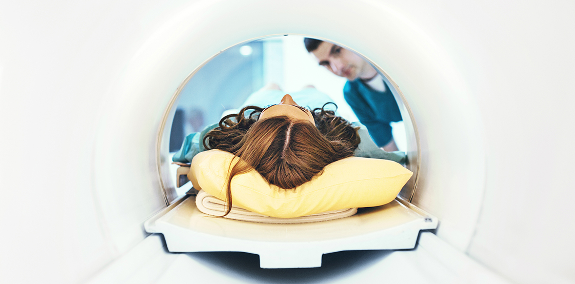
In the study, which was published late last year in the Annals of Rheumatic Diseases, around 800 healthy volunteers aged over 45 underwent an MRI scan.
The researchers expected that around 6-10 people (1%) in the study would have undiagnosed axial spondyloarthritis, based on earlier community data.
But, when the MRI results came back, the number of people with scans suggestive of axial spondyloarthritis was much higher, suggesting a problem with the interpretation of the images.
Structural sacroiliac joint bone marrow oedema, vertebral corners bone marrow oedema and post-inflammatory fatty lesions were seen in 17%, 28% and 81% of volunteers, respectively.
These abnormalities weren’t always just popping up once in each MRI scan, but several times in many patients, which would usually lead rheumatologists to a strong suspicion of axial spondyloarthritis.
The researchers determined whether the standard ASAS axial spondyloarthritis classification criteria would have been fulfilled by the MRI findings.
Their study reveals that this traditional way of interpreting MRI has a high false positive rate, if not evaluated in the context of clinical symptoms.
The researchers are now calling for caution around using a ‘positive MRI’ as proof that a patient has axial spondyloarthritis.
“These data suggest that the current definition of MRI changes in the structural sacroiliac joint used for the classification of axial spondyloarthritis need an update,” the authors said.
Professor Paul Bird, a rheumatologist based in Kogarah in NSW and the director of Optimus Clinical Research, “This is a very large study that confirms our suspicion regarding MRI reviewed in isolation.
“It is important for the clinicians to understand that the current criteria used for MRI interpretation must always be interpreted in the light of clinical findings rather than alone.
“The study shows that in a substantial proportion of patients, there was evidence of fat metaplasia and isolated areas of increased T2 signal intensity.
“As our understanding of MRI progresses, and more data becomes available, we will be able to be more specific about MRI findings and their relationship to active inflammatory disease. This study reinforces the need for more information regarding MRI in patients with lower back pain and in particular the group of patients who present with inflammatory low back pain.”
“In recent years, ASAS has focussed on validating the definition of positive MRI findings among patients with axial SpA,” said Dr Lionel Schachna, a rheumatologist based in Victoria.
“Recent reports suggest that changes of bone marrow oedema (BMO) may not be as specific for axial SpA as once thought. In previous studies, BMO on MRI was identified in up to 23% of patients with mechanical back pain and 7% of healthy volunteers. Therefore, the imaging arm of ASAS classification criteria is prone to false positive classification.”
On the basis of their findings, the authors suggested that five or more spinal lesions should be required to define a ‘positive spinal MRI’, said Dr Schachna.
“Importantly, the authors highlight the qualitative aspect of the interpretation of MRI changes particularly the specificity of deep lesions (defined as signal ≥1 cm from the articular surface).”
A major limitation of the study was the paucity of relevant clinical data, said Dr Schachna. “An accurate diagnosis of axial SpA still requires careful interpretation of the history and physical signs by an expert physician.”
