Doctors who use a biopsy to confirm temporal arteritis can now rely on a PET/CT scan for diagnosis, a study says
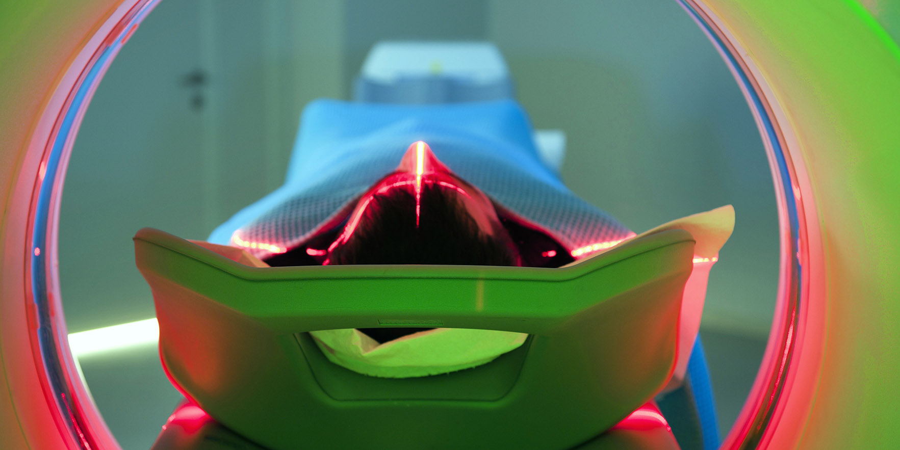
A new study recommends PET/CT as a first line diagnostic test for giant cell arteritis with biopsy often skipping affected vessels.
The study published this month in Arthritis and Rheumatology compared the diagnostic accuracy of biopsy, PET/CT and a six month follow up consultation with patients suspected of having temporal arteritis.
An accurate diagnosis is important with giant cell arteritis patients as the inflammatory disorder can cause permanent vision loss.
The author Dr Anthony Sammel, a rheumatologist at the Royal North Shore hospital in Sydney, said this is the first study to examine the accuracy of PET/CT technology scanning the head, neck and chest of patients suspected to have giant cell arteritis.
“We looked at the smaller arteries in the head and the neck. On older scanners, which were used in previous studies, they hadn’t been able to visualise those arteries,” he said.
As a result, doctors have historically ordered a biopsy to test for temporal arteritis, but Dr Sammel said the condition could be absent in the artery being tested.
“We think PET/CT should be a first line test. If the result is equivocal or doesn’t fit with your clinical suspicion, proceed to a biopsy.”
According to the study, up to 20% of patients with giant cell arteritis will have their diagnosis missed by a biopsy sample.
The study compared the accuracy of the different diagnostic methods across 64 patients who had experienced headache, a change in vision, fever or mastication pain.
The patients suspected of having temporal arteritis had undergone a c-reactive protein test (CRP) and an erythrocyte sedimentation rate (ESR) and were taking corticosteroids to reduce their risk of complications.
The scans and biopsy results were reported blindly, but the clinical history of the patient was known to the doctors involved in the diagnosis.
PET/CT had a sensitivity of 92% and specificity of 85% for the diagnosis of GCA when compared with temporal artery biopsy.
Only one of 12 biopsy positive patients had a negative scan. The sensitivity was 71% and specificity was 91% when PET/CT was compared with the six-month clinical diagnosis.
Dr Sammel said the study also showed using PET/CT was useful to confirm an alternate diagnosis for patients.
“Patients often come to us with high blood markers, but they can be high due to infection or cancer,” he said.
From using PET/CT 20% of patients received an alternative diagnosis including five cancers and seven infections.
But Dr Sammel also said PET/CT results should be married with the patients’ clinical picture.
“There will be a proportion of patients where either the PET scan result is not clear, or clinical suspicion is discordant with the PET scan result. In that case, I would go on to do a biopsy,” he said.
Arthritis and Rheumatology, 8 March 2019.


